Why Pre‑Pointe Assessments Matter for Young Dancers
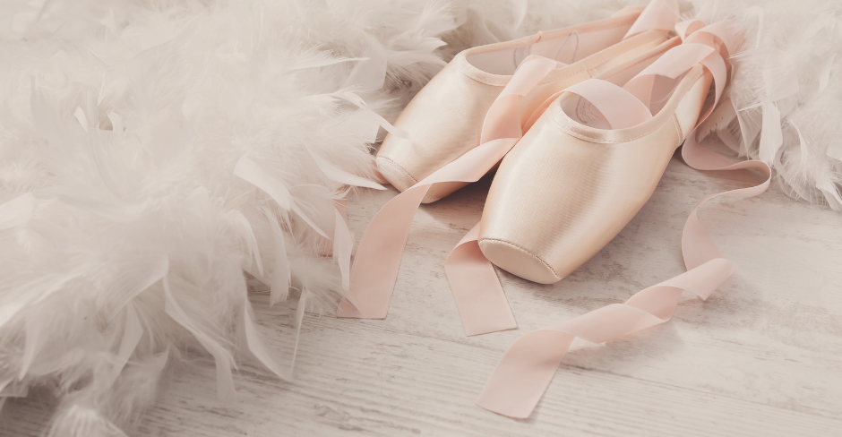


For many young dancers, the dream of going en pointe is a milestone filled with excitement, pride, and anticipation. But beneath the sparkle of new pointe shoes lies a crucial truth: pointe work places significant physical demands on a developing body. Ensuring a dancer is truly ready isn’t just about the skill required. It’s about safety, longevity, and setting them up for success.

That’s where Pre‑Pointe assessments come in. They are one of the most important steps a dancer can take on their journey, and they offer far more than a simple “yes” or “not yet.”

Preventative Work Is Always Better Than Rehabilitation

In dance, injuries often stem from overuse, poor technique, or progressing before the body is ready. Pointe work amplifies these risks. The feet, ankles, knees, hips, and spine must all work together under increased load and skill. Even small weaknesses can become big problems once a dancer is elevated onto the tips of their toes.

Identifying potential deficits in strength, mobility, alignment, or technique before a dancer goes en pointe dramatically reduces the likelihood of injury. Early intervention, identified with a Pre-Pointe assessment, means dancers can correct issues gradually, safely, and confidently. This avoids dealing with pain, frustration, or time away from the studio later on.

Pre‑Pointe assessments are designed exactly for this purpose: to protect the dancer’s body, support their development, and ensure their transition to pointe is both safe, sustainable and enjoyable.

Assessments Are Still Important for Dancers Already En Pointe Too

If a dancer is already en pointe, a Pre‑Pointe assessment is still highly valuable. It also doesn’t automatically mean they will be taken off pointe. The goal is not to stop their progress, but to ensure they are continuing en pointe as safely as possible. Even experienced dancers can develop strength imbalances, technique habits, or mobility limitations over time without realizing. An assessment helps identify any weaknesses that may place extra stress on the feet, ankles, or legs. Allowing us to address them early before they lead to injury is an important and healthy option to prolong and extend your pointe career. This proactive approach supports the dancer’s ongoing pointe journey, keeping them strong, confident, and performing at their best.

What Is a Pre‑Pointe Assessment?

A Pre‑Pointe assessment is a comprehensive evaluation performed by Jess, our dance‑trained clinician. It looks beyond the surface of technique and examines the physical foundations required for pointe work.

Assessments typically include:

  • Strength testing of the feet, ankles, calves, hips, and core
  • Flexibility and range‑of‑motion checks, especially through the ankles and big toe
  • Balance and proprioception tasks to assess control
  • Alignment and postural evaluation during classical technique
  • Functional movement tests specific to ballet (relevés, rises, pliés, jumps)
  • Technique analysis to identify habits that may affect pointe readiness

These assessments are often completed over several sessions. This staged approach allows dancers to focus on one element at a time, absorb feedback, and build the necessary foundations without feeling overwhelmed.

Why Pre‑Pointe Assessments Are So Important

1. They ensure the dancer is physically ready

Pointe work requires a level of strength, control, and mobility that cannot be rushed. Assessments highlight whether the dancer’s body is prepared for the demands ahead.

2. They prevent injury before it happens

By identifying areas needing improvement early, dancers can work on targeted exercises that reduce the risk of sprains, stress fractures, tendon issues, and overuse injuries.

3. They build confidence and clarity

Rather than guessing or relying solely on age or experience, dancers receive clear guidance on what they need to work on—and what they’re already doing well.

4. They support long‑term dance health

Bodies change as dancers grow. Regular reassessment ensures technique, strength, and mobility continue to progress safely, even after they’ve gone en pointe.

5. They reduce disappointment

Nothing is more disheartening for a young dancer than being told they aren’t ready. Assessments help avoid this by providing a structured pathway toward readiness, often starting 1–2 years before pointe work begins.


This Isn’t About Replacing the Dance Teacher’s Expertise

It’s important to emphasise that Pre‑Pointe assessments do not take away from the dance teacher’s expertise. Dance teachers are specialists in technique, artistry, musicality, and the day‑to‑day development of their students. Their role remains central. The assessment simply adds an additional, highly individualised layer of support—one that focuses on the dancer’s physical readiness in ways that aren’t always possible to address in a busy class environment. By combining the teacher’s technical guidance with a clinician’s understanding of strength, mobility, alignment, and injury prevention, dancers receive a more complete and personalized pathway toward pointe work. It’s a collaborative approach that surrounds the dancer with the combined support of both their teacher and their clinician.

A Strong Foundation Creates a Strong Dancer

Pre‑Pointe assessments aren’t just a box to tick—they’re an investment in a dancer’s future. They empower young dancers with knowledge, support, and a clear plan, ensuring their transition to pointe is safe, joyful, and sustainable.

With the right preparation, dancers don’t just go en pointe, they can THRIVE en pointe.

[Click Here] to book an appointment with our Dance Physiotherapist, Jess.


Written by Jessica Beer
Clinical Lead Physiotherapist | Dance Physiotherapy | Sports Physiotherapy

[Click Here] to read more about Jess


Additional Dance Content:


PhysioMotion Cairns
Shop 5 9-11 Stokes Street
Edmonton, Cairns QLD 4869

Servicing South Cairns Communities including Edmonton, Bentley Park, Mount Sheridan, White Rock, Bayview Heights, Woree and Gordonvale.

Herniated Discs: Should You Be Worried?

Back pain is one of the most common reasons people seek healthcare — and one diagnosis that often causes concern is a disc herniation.

But here’s the good news: despite how serious it may sound, most cases are highly treatable without surgery.

What is a Disc Herniation?

Your spine is made up of bones (vertebrae) separated by soft discs that act like cushions. These discs help absorb shock and allow movement.

A disc herniation happens when part of the inner material of the disc pushes outward, sometimes irritating nearby nerves. This can lead to symptoms such as:

  • Local back or neck pain.
  • Pain radiating into the arm or leg (like sciatica).
  • Numbness or tingling.
  • In some cases, muscle weakness.

Is it Serious?

Hearing that you have a “disc herniation” can be scary — but it’s important to put things into perspective.

Research shows that more than 90% of people recover well with conservative treatment, meaning surgery is usually not necessary.

In fact, many people have disc herniations and don’t even know it, because they have no symptoms at all.

What Helps?

During the early (acute) phase, your doctor may recommend medication to help manage pain and inflammation. This can be useful in the short term.

However, one of the most important parts of recovery is something simple — movement.

While rest might seem like the right choice, staying active (within your limits) is key to healing. Gentle movement helps:

  • Reduce stiffness
  • Improve circulation
  • Support tissue recovery
  • Restore confidence in movement

This is where physiotherapy plays a major role.

How Can Physiotherapy Help?

YES! We can guide you through a structured and safe recovery process, including:

  • Specific exercises to reduce pain and improve mobility.
  • Education about posture and daily habits.
  • Manual therapy techniques (when appropriate).
  • Gradual return to normal activities.

The goal is not just to reduce pain — but to help you move better and prevent future episodes.

Common Myths About Disc Herniation

“I need surgery to fix it.”
→ Most people recover without surgery.

“I should rest until the pain is gone.”
→ Too much rest can actually delay recovery.

“My spine is damaged forever.”
→ The body is very adaptable and capable of healing.

When Should You Seek Urgent Care?

Although rare, some symptoms require immediate medical attention, such as:

  • Loss of bladder or bowel control.
  • Severe or worsening weakness in the legs.
  • Numbness in the groin or saddle area.

If you experience any of these, seek medical help urgently.

Final Thoughts

A disc herniation may sound alarming, but in most cases, it is a manageable condition with excellent recovery outcomes.

With the right guidance, movement, and a tailored rehabilitation plan, you can return to your normal activities safely and confidently.

At PhysioMotion, we support you through every stage of your spine health — from acute pain to full recovery. Click here to book an appointment with one of our physiotherapists, and we will guide you towards the most appropriate treatment for your individual needs.


Written by:

Raul Loppi Goulart
Senior Physiotherapist

PhysioMotion Cairns
Servicing South Cairns including Edmonton, Bentley Park, Mount Sheridan, Gordonvale and surrounds

Understanding Pes Anserinus Bursitis: What It Is and How to Treat It????


If you’ve been feeling pain on the inside of your knee—especially when climbing stairs or getting up from a chair—you might be dealing with something called pes anserinus bursitis. It’s a common condition, and the good news is that it’s very treatable with the right care.

What Is Pes Anserinus?

The pes anserinus (Latin for “goose’s foot”) is where three muscles from your thigh (sartorius, gracilis, and semitendinosus) come together and attach to the inside of your shinbone, just below the knee. Underneath this area is a small fluid-filled sac called a bursa, which helps reduce friction between the muscles and bone.

When this bursa becomes irritated or inflamed, it causes pain and swelling. This is called pes anserinus bursitis.

What Causes It?

Pes anserinus bursitis can happen for a few reasons:

  • Doing repetitive movements like running or cycling
  • Having tight muscles in your thighs or hips
  • Having arthritis or poor posture
  • Getting hit or bumped on the inside of your knee

Common Symptoms

You might notice:

  • Pain on the inner side of your knee, a few centimeters below the joint
  • Tenderness when you press on the area
  • Swelling or warmth
  • Pain when going up stairs or standing from a seated position
  • No clicking or locking in the knee (which helps rule out other knee problems)
  • Pulling or tightness on the inside or back of the thigh 


How Physiotherapy Can Help

Physiotherapy is a great way to treat pes anserinus bursitis. Here’s what it usually involves:

Step 1: Reduce Pain and Swelling

  • Rest and avoid activities that make it worse
  • Use ice packs to calm inflammation
  • Gentle massage or hands-on therapy
  • Medication (like anti-inflammatories) if recommended by your doctor

Step 2: Stretch Tight Muscles to improve flexibility in your hips and legs

Step 3: Strengthen Key Muscles and practice safe exercise to support your knees

Step 4: Improve Movement and Posture

  • Learn better ways to walk, climb stairs, and move during daily tasks
  • Correct poor alignment or posture that may be causing strain

Step 5: Use Helpful Tools

  • Taping or bracing to support the knee
  • Orthotics (shoe inserts) if your foot posture is contributing


Pes anserinus bursitis can be frustrating, but with the right treatment plan, most people recover well. If you’re experiencing knee pain that fits this description, our dedicated team of physiotherapists can help you get back to moving comfortably and confidently.

Acceleration and Deceleration in Running & Sport: Why It Matters

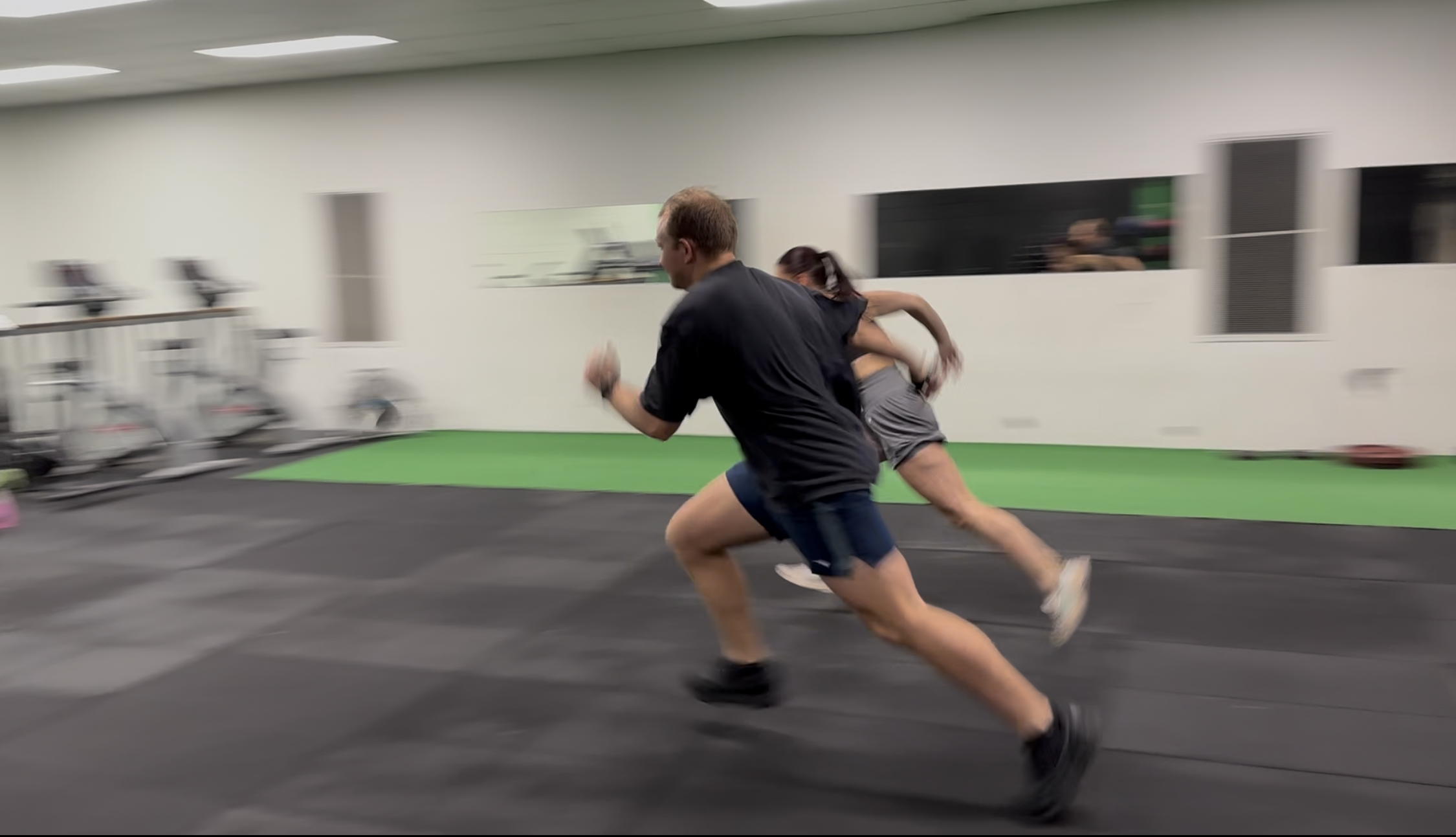
When we think about running, we focus on speed and endurance, but rarely do we pay attention to two critical phases: acceleration and deceleration. These components are essential not only for performance but for reducing the risk of injury. 


What Is Acceleration and Deceleration?

  • Acceleration is the process of increasing speed, like when you push off at the start of a run or sprint to chase a ball.
  • Deceleration is the ability to slow down or stop safely and efficiently. This is crucial when navigating turns, stopping quickly, or changing direction.

Both phases place high demands on your muscles, joints, and nervous system. They require strong coordination, muscle control, and balance—particularly through the hips, knees, and ankles.


Why It Matters for You

Many running-related injuries occur during deceleration, when the body absorbs high forces while trying to slow down. Poor control in these moments can lead to issues like knee pain, hamstring strains, or Achilles tendinopathy. Similarly, acceleration without proper strength and form can overload tissues, reduce efficiency and increase risk injury. 

When recovering from an injury, retraining acceleration and deceleration is a crucial step before safely returning to sport. These movements simulate the real demands of running and game play, where sudden starts, stops and change of directions are common.


How Physiotherapy Can Help

One of our physiotherapists can assess how well you accelerate and decelerate during running. Through movement analysis, strength training, and technique drills, we can help you:

  • Improve running efficiency
  • Reduce injury risk
  • Enhance performance in both straight-line running and change-of-direction sports
  • Safely return to sport


Written by


Levi Norsworthy
Physiotherapist


PhysioMotion Cairns
Shop 5/9-11 Stokes St
Edmonton QLD 4869

Fat Pad Impingement – Anterior Knee Pain

What is fat pad impingement?

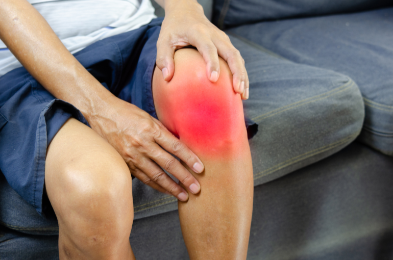
Do you get knee pain when going up or down stairs? Did you fall onto your knee one day and it’s still sore? What you may be experiencing is fat pad impingement, also referred to as Hoffa’s fat pad. Your fat pad is a pocket of soft tissue that sits underneath your knee cap at the front of your knee. It acts as a cushion between your knee cap (patella) and your shin bone (tibia). Sometimes this fat pad can be compressed causing what we call fat pad impingement. If the fat pad becomes inflamed, this can cause scar tissue to form. This scar tissue can lead to less room around the soft tissue of the knees causing further impingement when you try and straighten your knee. 

How can you injure your fat pad?

Injuries to your fat pad can be caused by a variety of injuries:

  • Tight quadriceps (thigh muscles)
  • Fall onto the front of the knee or a direct hit to the front of the knee
  • Overloading of the knee e.g. frequent going up/down stairs
  • An injury where the knee is pushed into hyper-extension 

Why does it hurt so much?

If you are wondering why your knee hurts so much – it very well could be the fat pad. The fat pad is a highly innervated and vascularized structure which means it has lots of nerve endings and blood vessels that supply it. This causes increased pain levels as there are lots of nerves in the area. 

Who may suffer from fat pad injuries?

  • Higher level sports where a blow to the knee may occur e.g. rugby, martial arts 
  • People who have experienced past injuries e.g. ACL tear
  • People who have weakness around the hip or knee
  • People who have a history of hyper-extension in the knee  
  • More common in young women
  • Those that partake in jumping sports 

Symptoms

  • Pain when straightening the knee especially towards the end of the movement 
  • Pain with being on your feet for hours
  • Difficulty doing up/down stairs 
  • Inflammation and swelling under the knee cap
  • Burning or tingling under the knee cap 
  • Stiffness in the knee and reduced mobility
  • May have difficulty walking or moving the knee if irritable 

Management

  • Ice 
  • Avoid aggravating activities – this may require a short break from sports
  • Braces for the knee 
  • Hands on therapy
  • Exercise for stretching and strengthening 
  • Taping 

Exercises to try at home

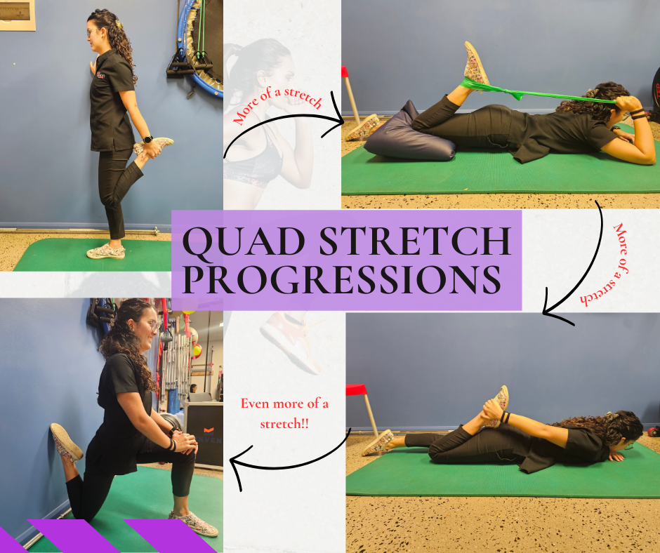
  • Quads stretching
  • Knee extension holds against a wall or with a resistance band if irritable. Aim to hold this position for 30 seconds. You may feel some discomfort during this exercise but it should not make your knee worse afterwards. 
  • Heel slides to increase bending and straightening movement – don’t push the knee into extension if it is sore 

Golfer’s Elbow

Do you have forearm pain with wrist movement, gripping, or throwing? Does it travel down the inside of your forearm toward your wrist? You may have an elbow condition called medial epicondylitis, or commonly referred to as Golfer’s Elbow.

Despite the name, 90% of cases are not sport-related. Occupations with forceful or repetitive activities (such as carpentry, plumbing, and construction) are also at risk of developing this issue due to the movements required for the job. Unsurprisingly, 3 out of 4 cases are in the dominant arm.

Medial epicondylitis, is classified as a tendinopathy and is commonly an overuse injury. This means that repetitive movements have caused an increase in loading of some key muscles (wrist flexors, pronator teres) which have caused changes, and an accumulation of microtears, within the tendon. It is possible for an acute trauma (sudden violent contraction of the muscles or a blow to the area) to cause this as well, but much less common.

Some common symptoms that are associated with Golfer’s Elbow are:
1. Pain from the inside of your elbow and down to your wrist.
2. Pain or weakness when throwing, holding, or squeezing objects.
3. Pain with forearm motion.
4. Elbow stiffness, weakness, or tingling in the ulnar nerve distribution (up to 20% of people can experience this).
5. Swelling, erythema, warmth (less likely with chronic).

How can we diagnose Golfer’s Elbow?
● MRI is ideal but generally used to rule out other causes of elbow pain.
● US is a quick, easy, and cost-effective way to assess muscles and tendons.
● Tenderness over the muscle tendon near where it attaches to the bone.
● Pain with muscle strength testing.
● Specific Golfer’s Elbow tests your Physiotherapist can perform which are considered positive if you experience pain on these test.
● Tinel’s test to assess for any nerve involvement.
● Valgus test to assess elbow ligaments particularly in throwing athletes.

Who is most likely to experience Golfer’s Elbow?
● 0.4% of the population experience Golfer’s Elbow.
● Usually people aged between 45-64 years old.
● More common in women than men.
● Labour intensive occupations with forceful, repetitive activities such as carpentry, plumbing, and construction work.
● Prevalence of up to 3.8% to 8.2% in some occupations.

Will Golfer’s Elbow recover?
People who experience Golfer’s Elbow can expect to make a full recovery and return to work or sport with appropriate treatment and advice.

What is the best treatment for Golfer’s Elbow?
Physiotherapy treatment is ideal. We have the ability to assess and treat the symptoms associated with Golfer’s Elbow and provide the best management plan for each person considering their work, sport and lifestyle goals.

Physiotherapy aims to achieve full, pain-free movement of the elbow and wrist and includes a strengthening program to return to usual activity. Hands on treatment is beneficial for reducing symptoms and can include dry needling, soft tissue releases and joint manipulation techniques.

In some cases night splinting, counterforce bracing or elbow taping may also be useful.

If you or someone you know are experiencing pain or discomfort over your elbow, contact one of our friendly Team Members today!


Ref: Kiel J, Kaiser K. Golfers Elbow. [Updated 2023 Jun 26]. In: StatPearls [Internet]. Treasure Island (FL): StatPearls Publishing; 2024 Jan-. Available from: https://www.ncbi.nlm.nih.gov/books/NBK519000/


Written by


Kalani McKenzie Tonga
Physiotherapist

PhysioMotion Cairns
Shop 5/9-11 Stokes Street
Edmonton QLD 4869

Piriformis Syndrome

Piriformis Syndrome

Piriformis syndrome is a condition which can cause pain, tingling, or numbness in the buttock and down the leg, which can often be mistaken for sciatica. The condition involves the piriformis muscle which is a muscle located deep in the buttock region and the closely located sciatic nerve.

The pain associated with piriformis syndrome is primarily located in the buttock region, with the potential to radiate down the back of the leg, following the distribution of the sciatic nerve. Typically the symptoms affect one side of the body more than the other and is often described as a deep ache.

Piriformis syndrome can affect individuals across various age groups, but it is more common in adults aged 30 to 40. It is slightly more common in women than men and can be more prevalent in active adults.

Certain activities can exacerbate the symptoms of piriformis syndrome:
Prolonged Sitting: Extended periods of sitting, especially on hard surfaces
Physical Activities: Running, walking, climbing stairs and standing

What can you do to help?

Some strategies can help alleviate the discomfort caused by piriformis syndrome:
Stretching Exercises: Regular, gentle stretching of the piriformis muscle and the surrounding muscles can reduce tightness and relieve pressure on the sciatic nerve.
Heat Therapy: Applying heat to the affected area can relax the muscle and reduce pain.
Avoid hard surfaces: Avoiding sitting on hard surfaces can prevent irritation of the piriformis muscle.
Self massage: Releasing the buttock and hip region including the piriformis muscle with massage or trigger point therapy (spikey ball).

Piriformis syndrome is a condition that can significantly impact daily activities and quality of life. By understanding the nature of the pain, identifying activities that aggravate symptoms, and implementing strategies to ease discomfort, individuals can manage this condition more effectively. If you suspect you might have piriformis syndrome, consulting with one of our physiotherapists is crucial for a proper diagnosis and to explore treatment options tailored to your specific needs.

Written By:

Levi Norsworthy
Physiotherapist

PhysioMotion Cairns
Servicing South Cairns including Edmonton, Bentley Park, Mount Sheridan, Gordonvale, Wright’s Creek and White Rock.

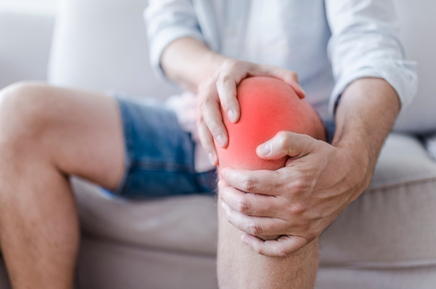
Patellofemoral (AKA Knee Cap) Pain


Do you ever get sore knees during daily activities, exercise or while you climb the stairs? Well if you do, you’re not alone. Knee pain with activity is a common concern reported to physiotherapists. Below is some information regarding patellofemoral joint pain and some advice on how you can help manage your symptoms at home. 


How Does the Knee Work:

The knee is a hinge joint which allows for bending and straightening movement. The patellofemoral joint is the joint between the kneecap (patella) and the intercondylar notch of the femur. This is where the kneecap travels during your bending movements. The tracking of the patella is influenced by the balanced pulling forces of the muscles around the thigh. 


The cause of PFJ knee pain: 

Patella femoral joint pain is a term used to describe symptoms underneath or around the knee cap. It can affect a wide range of people and is often aggravated with activities such as walking, running, up and down stairs, and squatting. As the structures on the inside and outside of the knee cap guide it during activity, this balance can be altered causing pain around the knee. This can be caused by the following:

  • Outside muscles being too tight
  • Inside structures being weaker than the outside muscles  
  • Excessive loads through the knee joint 
  • A combination of these factors

2 Ways to Help Your Pain:

1. Stretching:

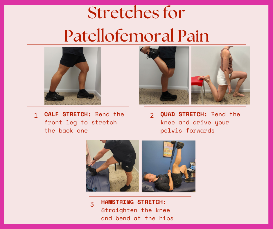
Stretching is a great way to reduce the tightness which can be contributing to your symptoms. Stretching regularly throughout the day and both before and after exercise can help. You should trial these stretches at home 

  • Quad 
    • While standing, raise your foot towards your glute. You should feel the stretch along the front of your thigh. Hold for 20 seconds and repeat three times at a mild to moderate stretch pain free.
  • HS
    • While sitting on the floor, have one leg out straight. Lean forward at your hips until you feel a stretch in the back of your thigh / knee. Hold for 20 seconds and repeat three times at a mild to moderate stretch pain free.
  • Calf 
    • Stand with one foot in front of the other facing a wall. Keep your back leg straight while leaning into the wall. You should feel a stretch in the back of your leg. Hold for 20 seconds and repeat three times at a mild to moderate stretch pain free.


2. Strengthening:

Strengthening is a greater way to maintain the balance within the knee. Often the reason muscles get tight is due to weakness (tired muscles get tight), so strengthening is a way to get you moving in the right direction. Trial these strengthening exercises at home:

  • Knee holds
    • Lay on your back and straighten your knee. Now try and push your knee into the bed and hold for 10 seconds repeat three times. 
  • Knee extensions 
    • While sitting on a chair, raise your foot so your knee is completely straight. Then lower your foot back to the floor. Repeated for 10 repetitions, three times.  
  • Sit to stands
    • From a seated position, raise to a standing position. Then lower yourself back into the chair. Repeated for 10 repetitions, three times.


Written By

Levi Norsworthy
Physiotherapist


PhysioMotion Cairns
Shop 5/9-11 Stokes Street
Edmonton QLD 4869

(Servicing Edmonton, Gordonvale, Bentley Park, Mount Sheridan Community)